
Glaucoma is often called the silent thief of sight — it steals your vision gradually, with no pain and often no early warning signs. Glaucoma screening is the single most reliable way to catch it before permanent damage occurs.
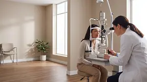
Glaucoma Screening: What to Expect and Who Needs It
What Is Glaucoma?
Glaucoma is a group of eye diseases that damage the optic nerve — the bundle of fibers that carries visual signals from your eye to your brain. Most forms of glaucoma are linked to elevated intraocular pressure (IOP), the fluid pressure inside the eye. When that pressure stays too high for too long, it gradually destroys optic nerve fibers.
What makes glaucoma so dangerous is that most people don't notice any symptoms until significant vision has already been lost. By the time side (peripheral) vision starts to fade noticeably, up to 40 percent of nerve fibers may already be gone. That lost vision cannot be restored.
The good news: when glaucoma is detected early through regular comprehensive eye exams, it can almost always be managed successfully with eye drops, laser therapy, or minimally invasive procedures — preserving vision for life.
Who Is at Risk for Glaucoma?
While glaucoma can develop in anyone, certain factors raise your risk substantially. Understanding your personal risk profile helps you and your eye doctor decide how often you should be screened.
High-risk groups include:
- People over 60 — Risk increases with age; adults over 60 are six times more likely to develop glaucoma.
- Family history — Having a first-degree relative (parent or sibling) with glaucoma raises your risk fourfold.
- High intraocular pressure — Elevated eye pressure is the most treatable risk factor; early detection allows intervention before nerve damage begins.
- African, Asian, or Hispanic heritage — These groups face higher rates of certain glaucoma subtypes, including angle-closure glaucoma.
- Diabetes and high blood pressure — Systemic vascular conditions can affect the blood supply to the optic nerve.
- Extreme nearsightedness or farsightedness — Highly myopic or hyperopic eyes have structural characteristics that elevate risk.
- Past eye injury or surgery — Trauma can alter fluid drainage in ways that raise pressure years later.

What Happens During a Glaucoma Screening?
A comprehensive glaucoma evaluation is painless, takes only a few minutes, and requires no preparation on your part. It is typically performed as part of a routine annual eye exam. Here is what the process looks like:
Tonometry — Measuring Eye Pressure
This is the most familiar glaucoma test: your doctor measures your intraocular pressure (IOP). The non-contact version uses a brief puff of air; the contact version (Goldmann applanation tonometry) lightly touches the eye surface with a tiny probe after numbing drops. Normal IOP falls between 10 and 21 mmHg. Elevated pressure alone does not confirm glaucoma, but it is a key red flag.
Ophthalmoscopy — Examining the Optic Nerve
Your doctor uses a bright light (and often dilating drops) to examine the optic nerve directly. A larger than normal "cup" relative to the disc — called a high cup-to-disc ratio — suggests possible nerve damage and warrants further testing.
Visual Field Test (Perimetry)
This computerized test maps your side (peripheral) vision. You press a button each time you see a small light flash in your visual field. Gaps or blank spots in the map can indicate nerve damage caused by glaucoma.
Pachymetry — Corneal Thickness
Corneal thickness affects how accurately tonometry reads true eye pressure. Thin corneas tend to underestimate IOP; thick corneas can overestimate it. A quick ultrasound or optical scan calibrates your pressure readings.
Gonioscopy and OCT Imaging
For high-risk patients, your doctor may also perform gonioscopy (examining the drainage angle with a special lens) or optical coherence tomography (OCT), which produces a detailed cross-section image of the optic nerve fiber layer — sometimes detecting subtle thinning years before visual field changes appear.
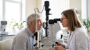
Key Glaucoma Risk Factors at a Glance
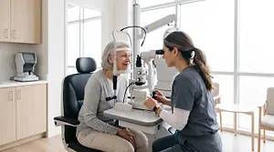
Age 60+
Risk rises significantly after 60. Adults in this age group are six times more likely to develop glaucoma, making annual screening essential.

Family History
A parent or sibling with glaucoma multiplies your personal risk fourfold. Genetic predisposition is one of the strongest predictors of this condition.
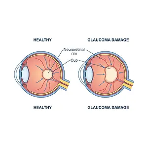
Elevated Eye Pressure
High intraocular pressure is the most treatable risk factor. Early detection via tonometry allows your doctor to start treatment before optic nerve damage begins.
How Often Should You Be Screened?
Screening frequency depends on your age, risk profile, and your eye doctor's findings. General guidelines from the American Academy of Ophthalmology suggest:
- Under 40, no risk factors: Every 5–10 years
- 40–54, no risk factors: Every 2–4 years
- 55–64, no risk factors: Every 1–3 years
- 65 and older: Every 1–2 years
- Any age with risk factors: Annually, or as directed by your eye doctor
These are minimum recommendations. If your eye doctor identifies optic nerve changes, elevated pressure, or visual field abnormalities, you may need more frequent monitoring — every 6 months or even more often. Regular follow-up is not optional; it is how glaucoma management works.
If you have been diagnosed with an eye condition that raises your glaucoma risk, such as pigment dispersion syndrome or pseudoexfoliation syndrome, your doctor will tailor a monitoring schedule to your individual situation.
What Happens If Glaucoma Is Found?
A positive glaucoma screening result — elevated pressure, suspicious optic nerve, or abnormal visual field — does not necessarily mean you have glaucoma. It means further evaluation is needed. Your eye doctor may repeat the tests, refer you to a glaucoma specialist, or schedule closer monitoring.
If glaucoma is confirmed, modern treatment options are effective and often straightforward:
- Prescription eye drops — The most common first-line treatment, these either reduce fluid production or improve drainage to lower IOP.
- Laser therapy (SLT) — Selective laser trabeculoplasty opens drainage channels in about 15 minutes, often reducing the need for daily eye drops.
- Minimally invasive glaucoma surgery (MIGS) — Tiny implants or micro-incisions improve fluid outflow with faster recovery than traditional surgery.
- Traditional glaucoma surgery — Reserved for advanced cases where other treatments have not lowered pressure enough.
The critical message: treatment works when started early. Eyes lost to advanced glaucoma cannot be restored. The investment of one annual eye exam is the most effective glaucoma prevention strategy available.
Frequently Asked Questions About Glaucoma Screening
Everyone should have their eyes checked for glaucoma, but those with the highest urgency are adults over 60, anyone with a family history of glaucoma, people with elevated eye pressure, and those with diabetes or high blood pressure. African Americans and Hispanic adults are also at higher risk and benefit from earlier and more frequent screening, starting around age 40.
Glaucoma screening is a series of painless tests performed during an eye exam to check for signs of the disease. It typically includes tonometry (measuring eye pressure), an optic nerve exam, and a visual field test. Advanced screenings may also include OCT imaging, which can detect nerve fiber thinning years before symptoms appear.
A standard glaucoma screening consists of at least three tests: tonometry to measure intraocular pressure, ophthalmoscopy to examine the optic nerve head, and perimetry (visual field testing) to check for peripheral vision gaps. Your doctor may add pachymetry to measure corneal thickness or OCT imaging for a detailed nerve fiber layer analysis.
Glaucoma screening is done as part of a comprehensive eye exam in your doctor's office. You'll sit in front of a slit lamp for the optic nerve exam, briefly hold your eyes open for the tonometry reading, and look at a screen for the visual field test. None of the tests require needles or incisions, and most are completed within 10 to 15 minutes total.
Medicare Part B covers one glaucoma screening per year for beneficiaries at high risk — including those with diabetes, a family history of glaucoma, African Americans over 50, and Hispanic Americans over 65. The test must be performed or supervised by an eye doctor or ophthalmologist. If you are unsure whether you qualify, ask our front desk team to help verify your coverage before your appointment.